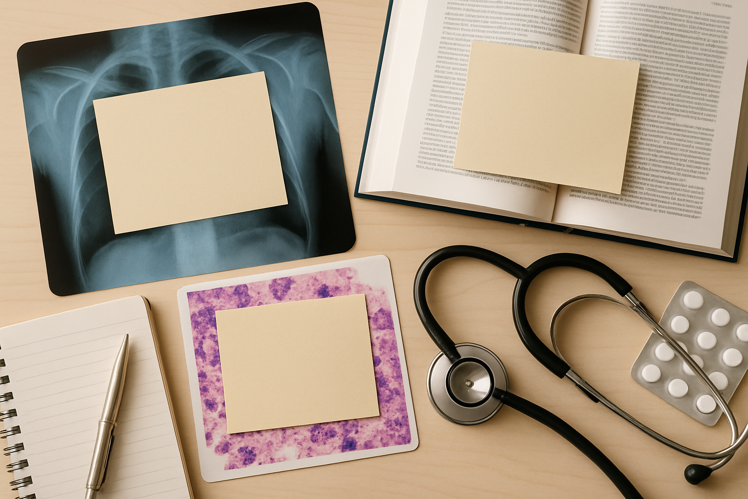
Why Image Occlusion Is Worth the Trouble
Visual recall drives clinical judgement. Recognising a pneumothorax on a chest film or a Reed–Sternberg cell on a biopsy slide depends on spotting spatial relationships, not memorising text descriptions.
Image occlusion turns passive image review into directed retrieval practice. You blank out the structure name and force your brain to label what you see. The first few hundred occlusions are slow, but when built efficiently, they become powerful visual anchors for diagnosis.
The problem: most students build them inefficiently—too many clicks, no tagging, and scattered files. That’s fixable.
Start With the Right Source Image
When choosing an image, start with clarity and educational value, not aesthetics.
- Resolution: At least 1600 px wide; lower-resolution slides blur when zoomed and confuse recognition.
- Colour balance: Especially for histopathology; avoid images with tinted coverslips or digital artefacts.
- Annotation-free: Start from a clean version without arrows or text overlays. You’ll add controlled cues later.
- Representative view: Pick classic appearances—lobar pneumonia consolidation, osteosarcoma lacework, etc.—that mirror exam-style material.
Save originals in an Image Occlusion folder. Prefix filenames with the topic (e.g., Resp_CXR_Pneumothorax_1.jpg). The 30 seconds you spend organising now will save hours searching later.
Streamline the Image Occlusion Process
1. Identify What’s Worth Covering
In radiology, cover:
- Unique or diagnostic findings: air bronchograms, meniscus sign, Hampton’s hump
- Key anatomical landmarks: costophrenic angles, mediastinal contours
- Contrast patterns on CT or ultrasound
In pathology:
- Cell or tissue features distinguishing one diagnosis from another
- Stain-specific highlights (PAS‑positive material, AFB rods)
- Architectural patterns (glandular vs. sheet-like)
Don’t mask every vessel or every nucleus. The best decks emphasise pattern recognition, not label memorisation.
2. Use AI to Draft Masks
Modern AI tools can auto-suggest occlusion areas. Cardivate integrates with image segmentation APIs and local models so you can:
- Upload the slide.
- Let the model label possible structures (e.g., “left upper lobe opacity”, “keratin pearl”).
- Review suggestions, accept or refine.
This cuts manual masking time by 60–70%. It also helps you notice overlooked findings—particularly useful when learning to read CT slices or immunohistochemistry panels.
3. Apply Meaningful Labels Immediately
Once your occlusion cards are generated, label them right away. Don’t rely on future-you remembering what “Image 103” meant.
Follow a consistent naming convention:
- Radiology: Modality – View – Finding
- Example:
CXR – PA – Pneumothorax border
- Example:
- Pathology: Organ – Stain – Feature
- Example:
Liver – H&E – Councilman body
- Example:
Consistent labelling aligns with Cardivate’s tag search, letting you filter all “CXR–PA” cards or all “Gram stain” slides during focused reviews.
Build Efficient Occlusion Cards
Use Layered Occlusions for Progressive Disclosure
Start with broad structures, then move into detailed ones. For example, in a CT head:
- Mask the lateral ventricles.
- Mask the caudate nucleus.
- Mask the internal capsule.
Arrange them as steps, not separate cards. In Cardivate, the layer ordering tool lets you keep each mask on a different layer, unveiling them sequentially in review. This prevents you from memorising pixel-level cues and trains spatial integration.
Keep Each Card Clinically Focused
One card = one diagnostic idea. Avoid crowding multiple unrelated findings (e.g. cardiomegaly and pleural effusion) on one occlusion. Better to make two smaller cards that test each concept independently.
Use Contextual Hints, Not Giveaways
If two structures look similar (say, hepatic artery and bile duct), provide context via partial outlines or colour-coded borders instead of nearby labels. The brain latches onto spatial context first; small cues accelerate pattern encoding.
Apply AI Beyond Masking
Image occlusion isn’t just about drawing boxes. AI can accelerate tagging and explanation writing too.
Auto‑Tagging
Feed your labelled occlusions through an AI tagger that generates hierarchical tags like:
Modality: MRIRegion: BrainFinding: GlioblastomaKeyword: ring enhancement
Cardivate reads these as deck tags. You can later review only Finding:Glioblastoma cards or build filtered study sessions on Modality:MRI.
Draft Short Rationales
Autogenerate a two‑line explanation for each occlusion: what the feature represents and why it matters clinically.
Example skeleton:
“Lobar consolidation with air bronchograms indicates alveoli filled with exudate while bronchi remain air-filled, typical in bacterial pneumonia.”
Editing the AI draft into your own words deepens understanding. Don’t skip this—retrieval is stronger when the rationale is re‑phrased manually.
Tagging for Retrieval Practice
Once your occlusions are ready, tagging determines how effectively you’ll revisit them.
Tag dimensions that actually guide study sessions:
- Modality (CXR, CT, MRI, histology)
- System (respiratory, gastrointestinal, haematology)
- Finding type (mass, opacity, pattern)
- Difficulty (easy, review, challenging)
Avoid inconsistent tag names. “Neuro” and “neurology” aren’t treated the same. Stick to a fixed tag list—a short one you can remember. Investing an hour in tag structure pays off every exam cycle.
With Cardivate, use Tag Templates to apply tag sets automatically to new occlusion imports. That keeps decks uniform even when multiple classmates contribute.
Integrate with Daily Study Flow
Radiology and pathology visuals saturate every unit. To prevent overload:
Daily Quick Review
Schedule 10–15 minutes daily to review 10–20 occlusion cards. Spaced repetition will do the rest.
Add While Studying, Not Afterward
The most efficient time to build occlusion cards is immediately after covering a case or lecture—when the image is top of mind. Waiting “until later” doubles the effort because you must re‑locate and re‑understand each image.
Use Filters for Focused Sessions
Before exam periods, filter by:
Modality: CXRandFinding: opacityStain: GramandSystem: Microbiology
Targeted sessions condense hundreds of cards into pattern drills that match clinical questions.
Efficiency Metrics to Track
Without feedback, you can’t improve speed. Track:
- Masking time per image: aim for under 3 minutes after your first 20.
- Percentage auto-generated by AI: target > 60%.
- Review accuracy: monitor first‑pass recall vs. retention after 7 days.
- Tag completeness: > 95% of cards should have all major tag fields.
Use Cardivate’s Deck Stats dashboard or any spreadsheet to monitor progress. Treat it like lab data—you can’t optimise what you don’t measure.
Troubleshooting Common Pitfalls
Problem: Overlapping Masks Cause Confusion
Keep margins of a few pixels between masks. When features genuinely overlap (e.g. arteries and ducts), use semi‑transparent fills to distinguish.
Problem: AI Labels Incorrect Structures
Provide 3–5 seed hints manually. Segmentation models refine accuracy after minimal manual correction.
Problem: Deck Feels Disjointed
Group occlusions by region, not by lecture. Combine images that teach similar diagnostic reasoning (all “upper‑lobe opacities”) for spaced comparison.
Problem: Forgetting Clinical Relevance
Pair every visual occlusion with a one‑line clinical cue: “Typical in Mycoplasma infection.” That link moves image recall from rote pattern to applied recognition.
Maintain and Share Responsibly
Images often come from institutional resources or open atlases. Before sharing occlusion decks:
- Verify that base images are under open or educational licences.
- Strip patient identifiers from radiographs.
- Credit original sources in the note’s reference field.
A shared deck built on clean, legally safe material becomes a sustainable study repository for your entire cohort.
The Bottom Line
Efficient image occlusion sits at the intersection of repetition and reasoning. You’re training pattern recognition the same way radiologists and pathologists do—by controlled exposure and active recall.
Use AI to handle the grunt work: initial masks, tagging, and quick rationales. Your energy should go into decision practice—the moment your brain names what it sees. Build systematically, tag consistently, and review steadily. The hours you save in deck creation can then go where they belong: interpreting the next image with confidence.